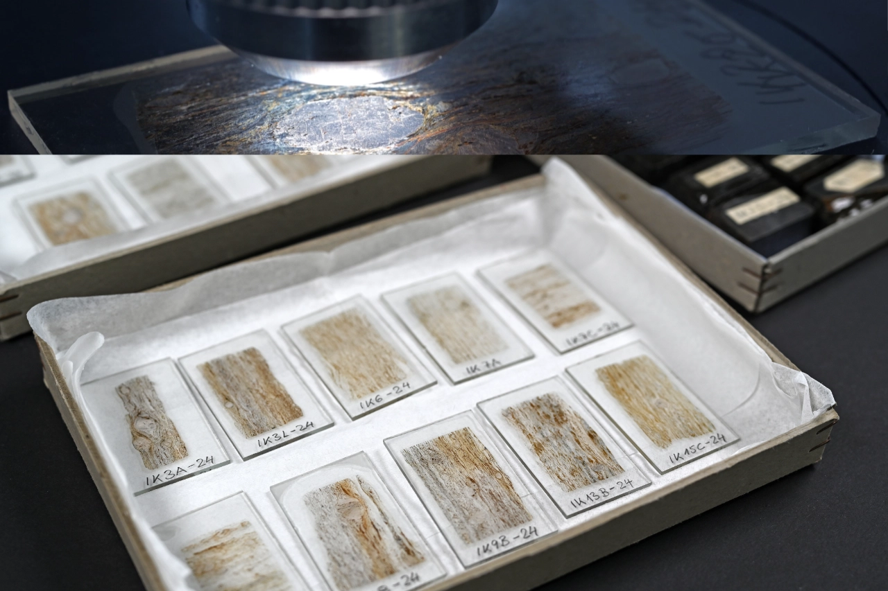
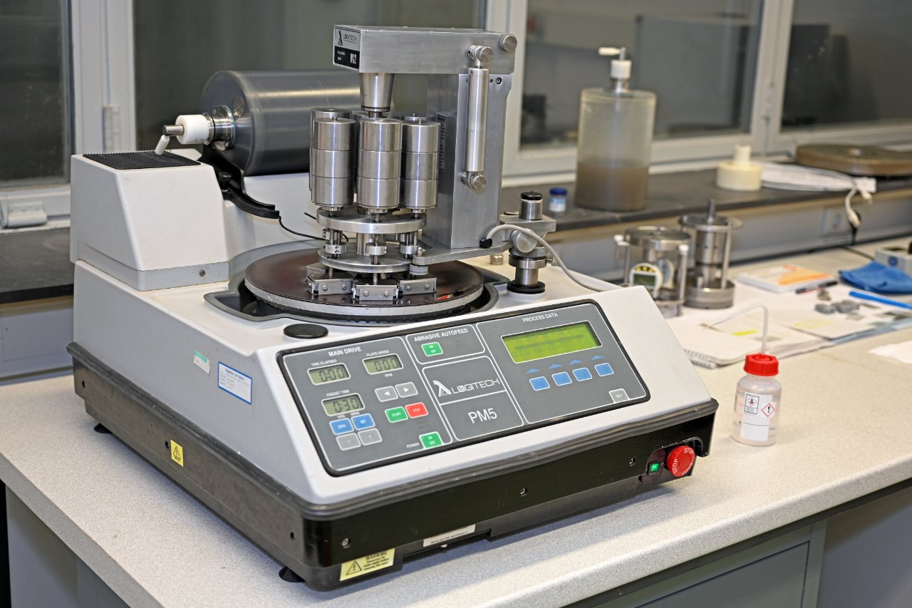
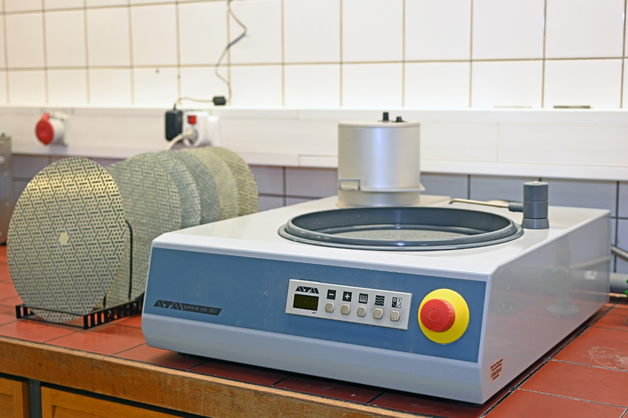
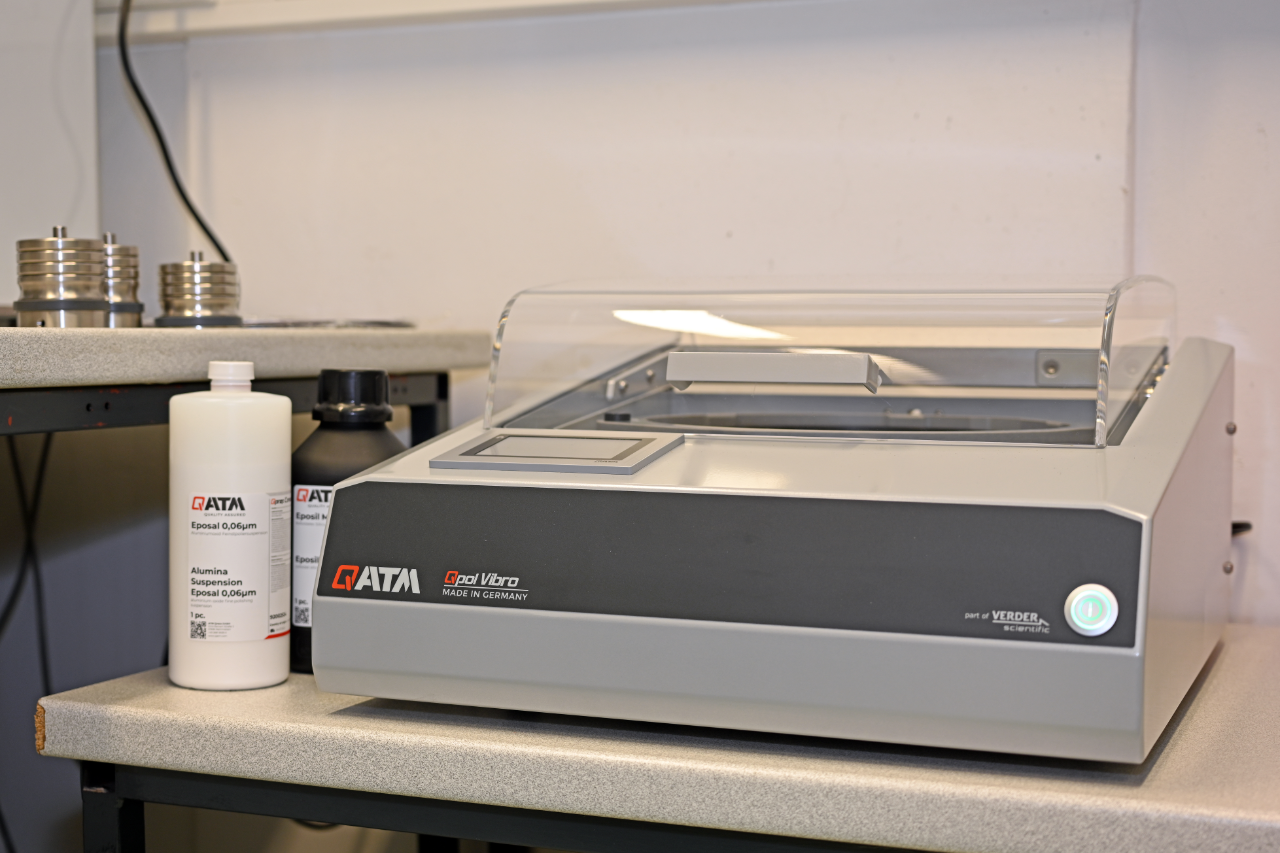
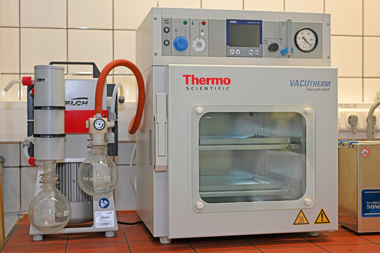
Production of rock thin sections and preparation of rock, mineral and fossil samples for further analysis.
Contact person: Prof. Dr. Evangelos Moulas – Research group Metamorphic Processes
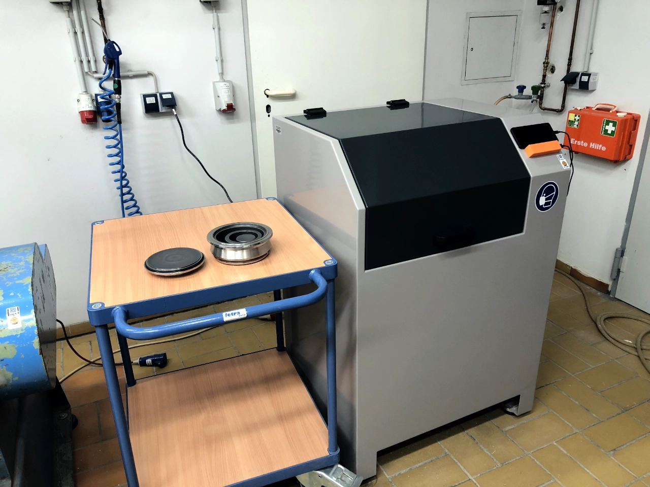
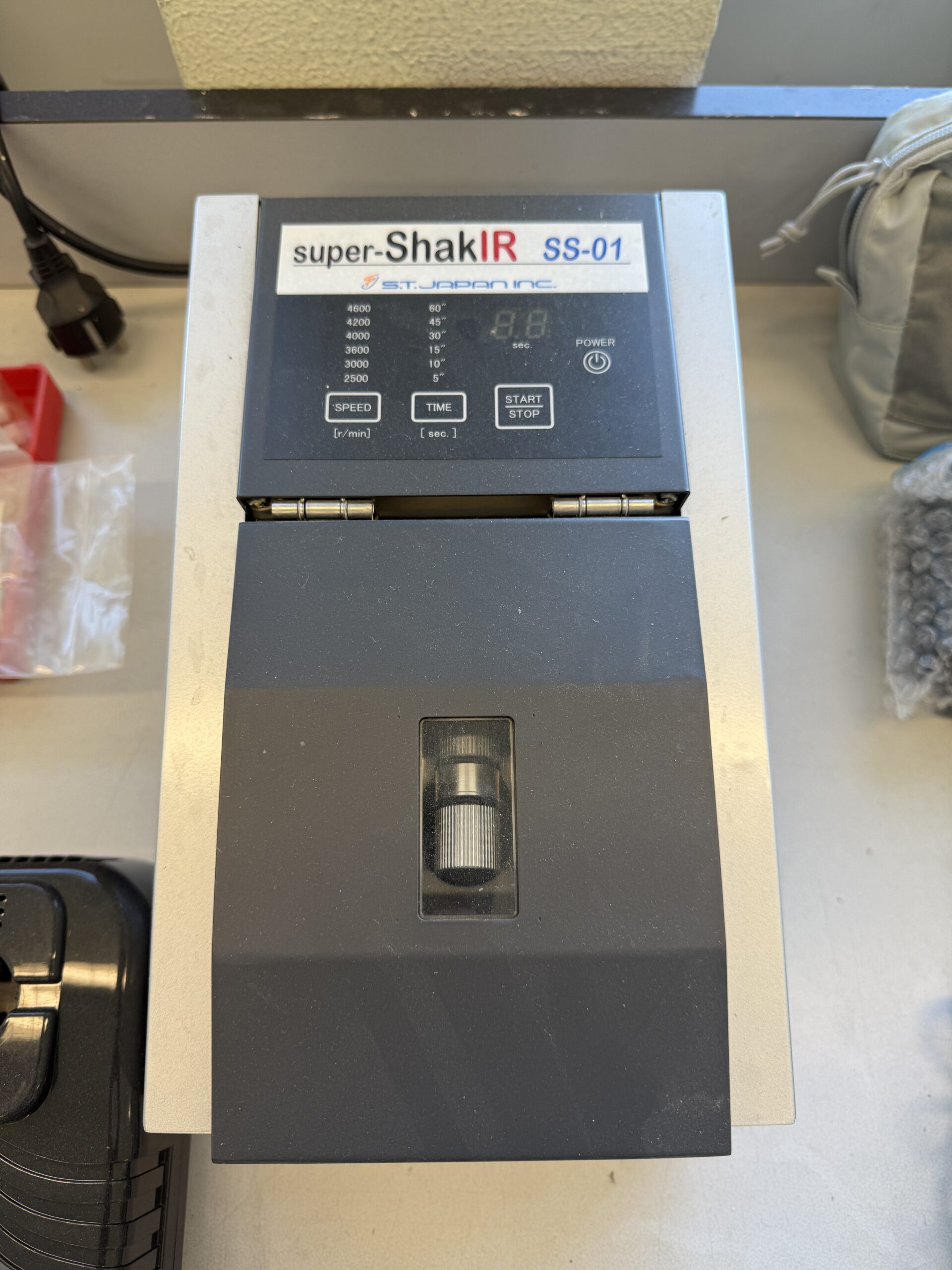
Disc vibration mill with tungsten carbide insert for processing crushed rock samples for further analytics such as X-ray fluorescence analysis (XRF) or X-ray diffractometry.
Hydraulic press, jaw crusher and roller mill for coarse and medium-fine crushing of rock samples
Additional equipment for sample preparation in various research groups:
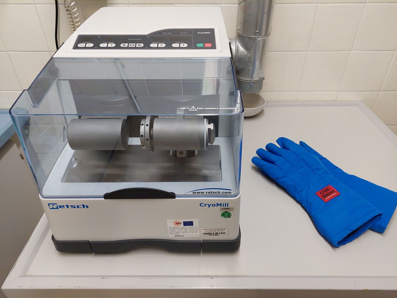
The Cryomill is a specialized ball mill used to gently grind soft tissue or biomineralizates to an analytical fineness while cooling the grinding jar and material with liquid nitrogen.
Contact person: Prof. Dr. Thomas Tütken – Research group Paleontology
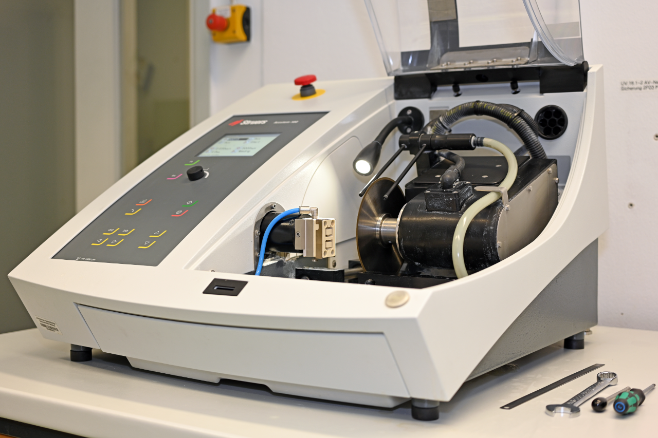
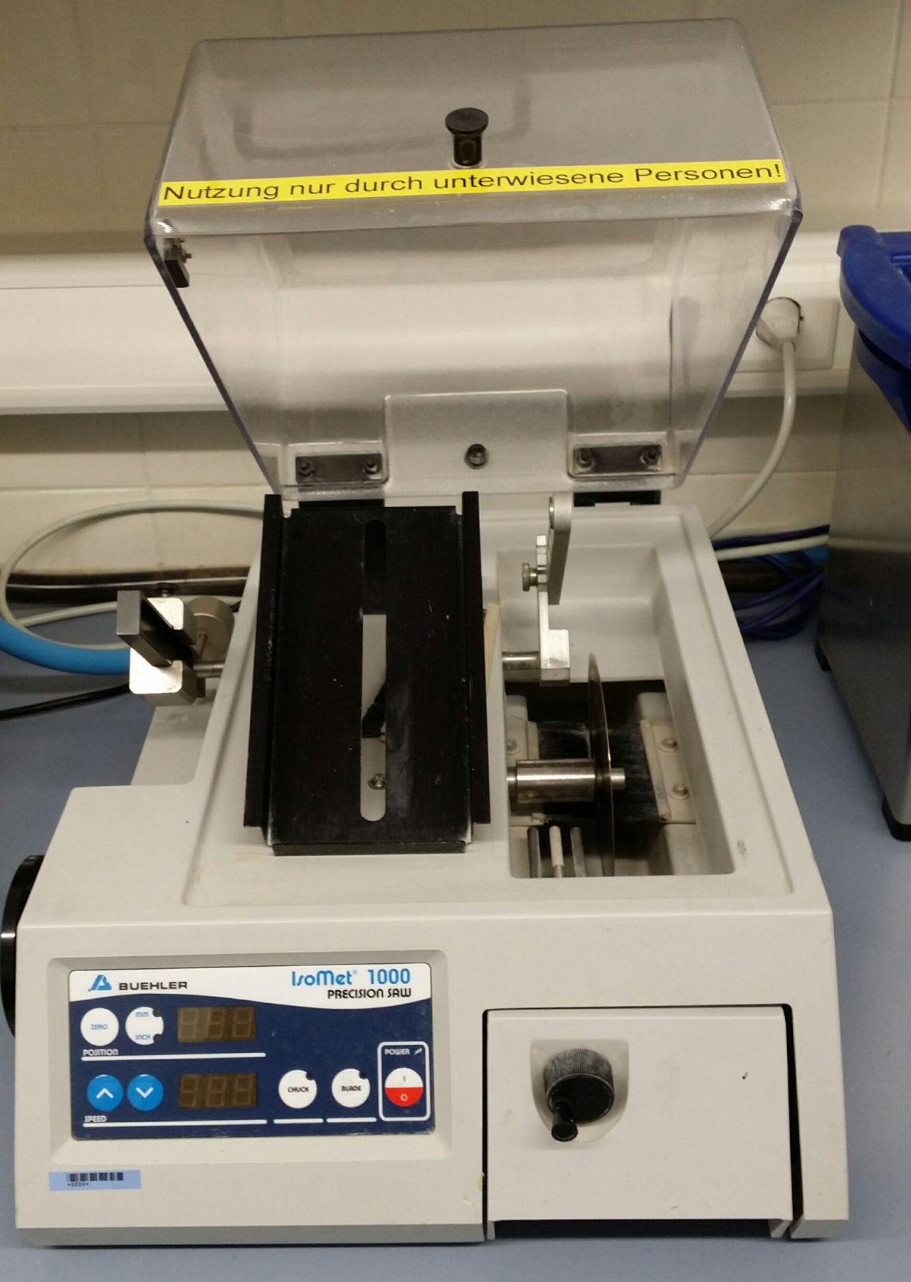
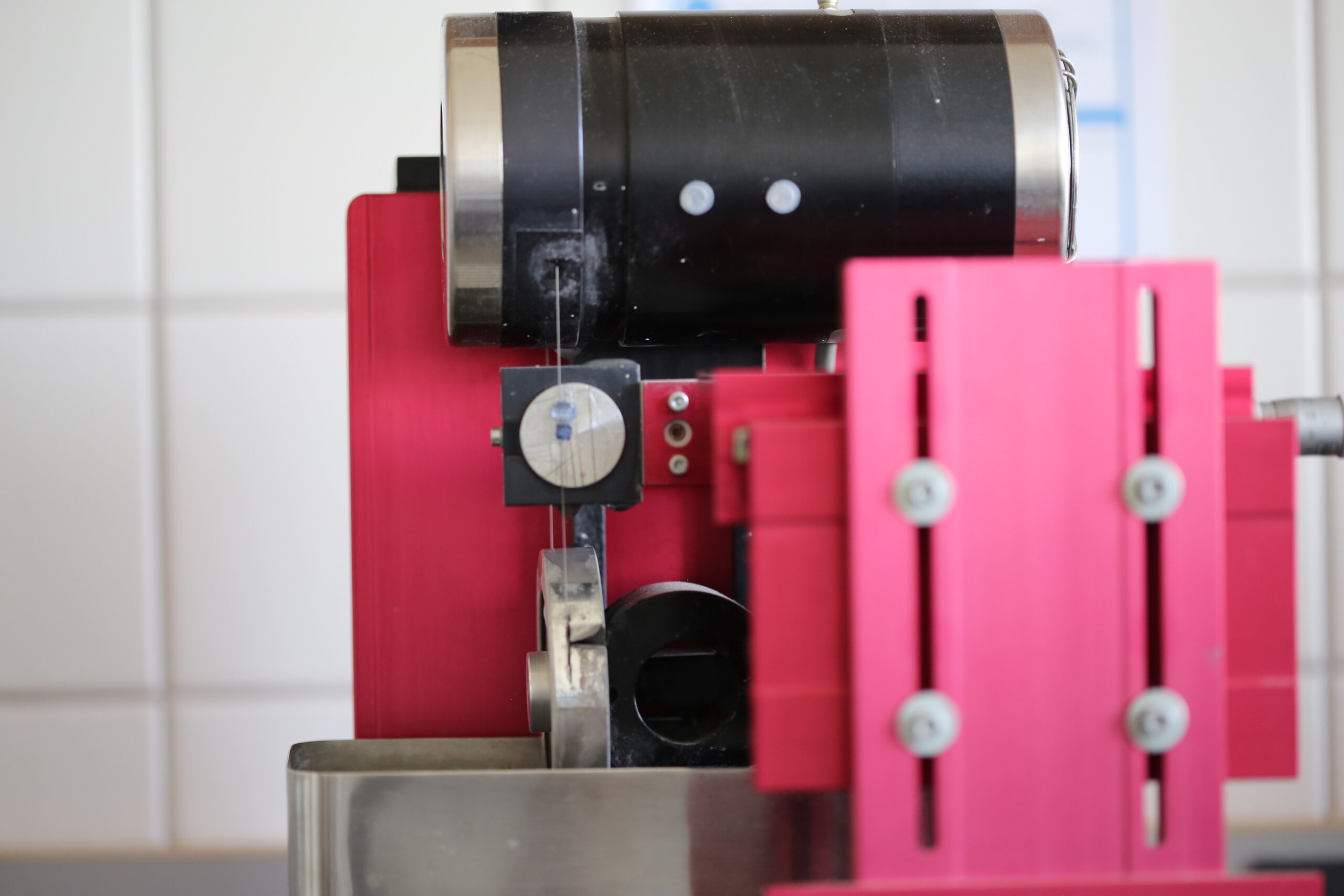
Used for precise, material-conserving cutting of smaller mineral, rock or hard-tissue samples with water-cooled diamond-coated saw blades and minimal sample loss.
Various research groups
Contact person: Dr. Tobias Häger – Research group Geomaterials and Gemstone Research
Contact person: Prof. Dr. J. Castro – Research group Volcanology
Contact person: Prof. Dr. D. Scholz – Research Group Speleothem Research
Production of homogeneous glasses from small quantities of rock powder.
Contact person: Prof. Dr. D. Scholz – Research Group Speleothem Research
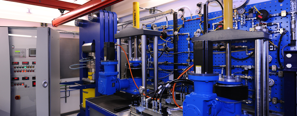
The Petrology work group has a piston-cylinder laboratory with two presses in a modified Boyd-England design. The equipment includes a 650-ton apparatus with a 1-inch piston diameter and a 250-ton press with a 0.5-inch piston diameter.
Both apparatus can generate pressures of up to 3.0 GPa (30,000 atmospheres) and reach temperatures of up to 1500 °C.
Contact person: Dr. S. Buhre – Research group Petrology
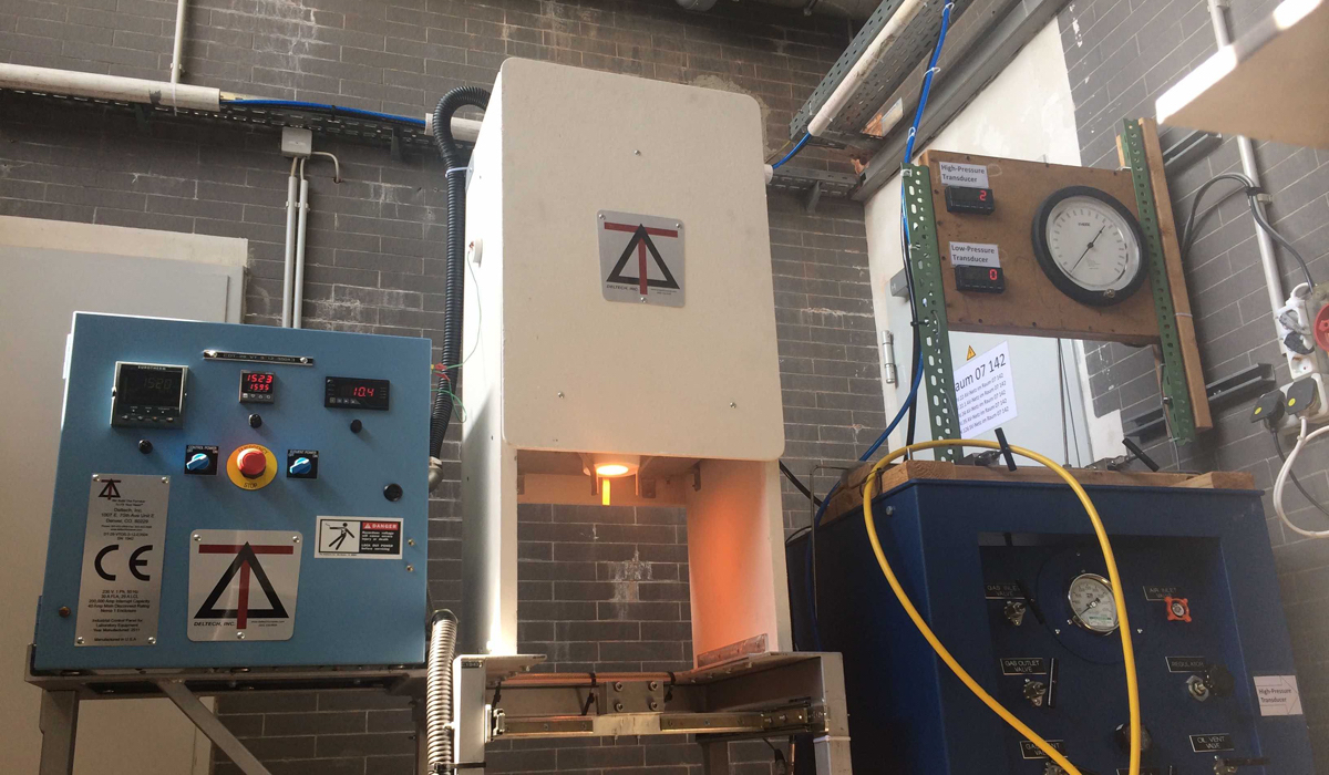
The multi-anvil apparatus can achieve pressures of up to 15 GPa (150,000 atmospheres) and temperatures of up to 2500°.
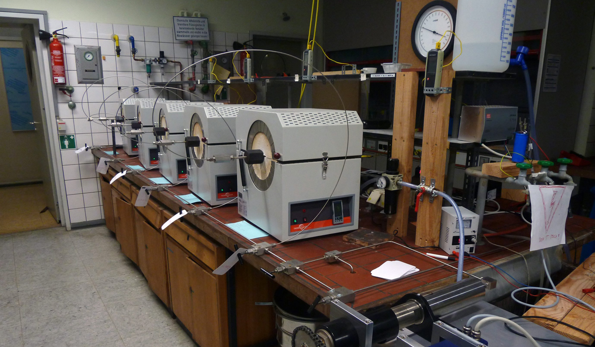
Hydrothermal autoclave system with Thermoconcept hinged tube furnaces
Contact person: Prof. Dr. J. Castro – Research group Volcanology
Contact person: Prof. Dr. J. Castro – Research group Volcanology
Contact person: Prof. Dr. J. Castro – Research group Volcanology
Contact person: Prof. Dr. J. Castro – Research group Volcanology
Our group conducts laboratory experiments on speleothem growth and the associated isotope fractionation using a setup that is unique worldwide. In a climate chamber, temperature, humidity, pCO2, the isotopic composition of atmospheric CO2, and drip water can be set and controlled. As in a natural cave, synthetic speleothems can be precipitated from dripping or flowing very thin solution films. This allows us to investigate the influence of various environmental parameters such as temperature and drip rate on the isotopic composition of speleothems under controlled conditions.
Contact person: Prof. Dr. Denis Scholz – Research group Speleothem Research
Laboratory experiments – climate chamber and Themo Scientific Delta Ray
(c) Peter Pulkowski
The electron microprobe (EMP) or electron probe micro-analyzer (EPMA) is based on a scanning electron microscope. However, it is equipped with 5 WDX spectrometers for quantitative analysis of major, minor, and trace element concentrations.
This allows the composition of the smallest areas to be analyzed non-destructively, meaning that even mineral grains measuring just a few micrometers can be analyzed in-situ (i.e., within the rock matrix) with high precision. In addition to rock samples, the smallest glass particles, gemstones, bones, teeth, skeletons of marine organisms, and much more can also be examined.
Electrons are emitted from a Schottky emitter. This is a special type of electron source based on thermally assisted field emission. It consists of a very finely tapered tungsten cathode coated with a small amount of zirconium oxide. This coating lowers the effective work function of the electrons at the metal surface.
The cathode is moderately heated and simultaneously exposed to a strong electric field. Through the combination of thermal energy and field emission, electrons can overcome the potential barrier at the tip and escape into the vacuum. The emission area is extremely small, resulting in very high beam density. Compared to conventional thermal electron sources, the Schottky emitter provides a particularly bright, stable, and low-energy electron beam. This enables high spatial resolution, good beam stability, and reproducible conditions, which are crucial for precise geochemical analyses in the electron microprobe.
The electrons thus generated are accelerated in a high vacuum by an electric potential of up to 30 kV toward the sample. On their way there, the charge carriers pass through a series of electromagnetic coils that shape and focus the beam before it strikes the sample. There, interaction occurs with the atoms of the different elements in the sample, which are excited to emit their specific X-ray radiation. The interaction volume is only a few cubic micrometers, depending on the acceleration voltage. The X-ray spectrometers function like X-ray filters and measure the number of X-ray pulses for a specific element sequentially (normalized to the measurement time and beam current). The measured intensities are then compared with measurements on reference materials (whose compositions have been well determined beforehand) and element concentrations are calculated from this. The matrix correction performed after the measurement is needed to correct for the mutual influence of the elements on one another.
In principle, the EMS can analyze elements from Be to Pu (see periodic table) in all solid materials, provided they are vacuum-stable and do not change under the influence of electron bombardment. For quantitative analyses, a very good surface polish is also mandatory. Non-conductive materials must be coated with a 15-20 nm thick carbon layer to prevent negative charging of the sample.
Two of the 5 wavelength-dispersive spectrometers operate with Ar/methane flow counters, which in combination with LDE1, LDEB, LDEC, TAP or TAPL, and PETL crystals are optimized for measuring light elements. The other spectrometers are equipped with Xe counters and a combination of PETL and LIFL crystals.
With the EDS system, it is possible to measure multiple elements simultaneously and thereby perform phase identification in a matter of seconds.
Contact person: Dr. S. Buhre – Research group Petrology
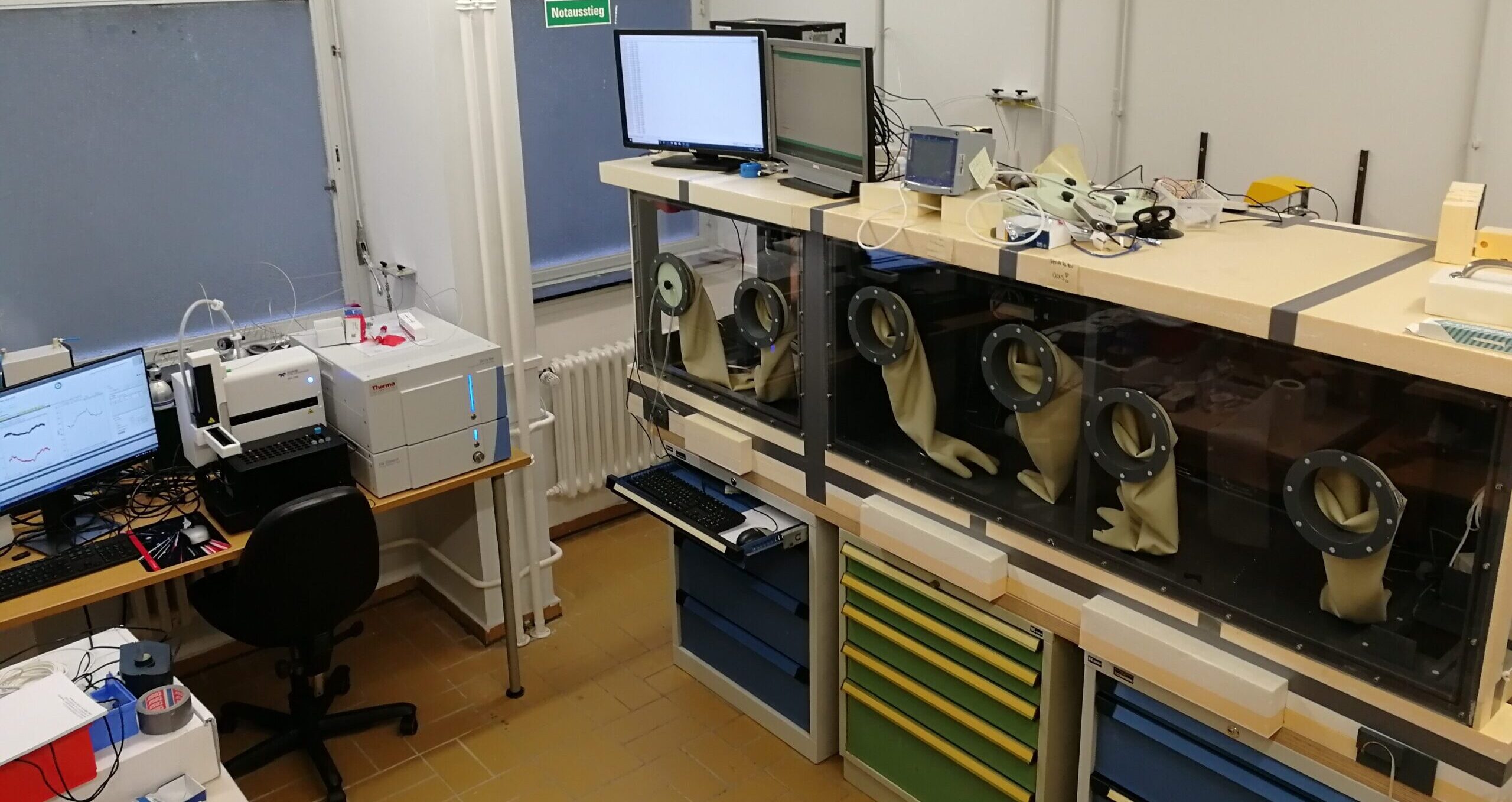
X-ray fluorescence analysis (XRF) is used for material analysis. It serves for the qualitative and quantitative determination of the elemental composition of solid, liquid, or pasty samples, such as: rocks, soils, minerals, clays, ores, steels, alloys, glasses, ceramics, building materials, slags, plastics, pastes, and oils.
XRF is used in research and in quality and production control in industry. Depending on the equipment of the analytical instrument, the determination of elements from atomic number 4 (Be) to 92 (U) is possible. The measurement range, depending on the element and the matrix, extends from 0.0001 wt% to 100 wt%.
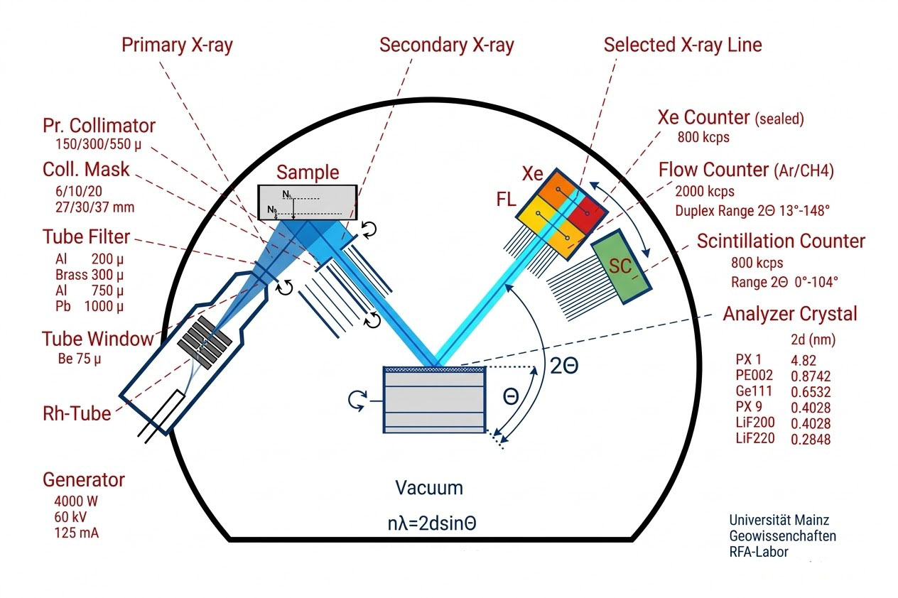
X-ray fluorescence analysis uses X-rays as primary radiation to excite a sample to be analyzed. Through interaction with the sample, secondary X-ray radiation (“fluorescence radiation”) is produced, which can be qualitatively and quantitatively determined after spectral decomposition by diffraction on a crystal. The wavelengths of this radiation are characteristic of the elements present in the sample. From the intensity of the characteristic radiation, the concentration of the elements to be determined can be calculated based on extensive correction and calibration programs.
In order to optimally address all questions, the spectrometer is equipped with various apertures, 3 collimators, 6 analyzer crystals (PX1, PE002, Ge111, PX9, LiF200, LiF220), and 3 detectors (flow, scintillation, and Xe proportional counters). This allows the best setting to be made for trace to major elements.
The main area of application at our institute is the determination of major and trace elements in geological samples. If required, special measurement programs (applications) are also developed. Semi-quantitative analyses of a wide range of solids can be carried out using the standardless IQ+ measurement procedure from Panalytical.
Contact person: Dipl.-Min. N. Groshopf – Research group Petrology
The Bruker M4 TORNADO is a powerful micro X-ray fluorescence (μXRF) system for non-destructive analysis of sediments, rocks and other materials. With a minimum spatial resolution of 20 µm, it enables the creation of high-resolution 2D maps of elemental distribution and spot analyses directly on the sample.
The system offers high measurement speed, ease of use, and flexible customization options for different sample formats and analysis types.
Contact person: Jun.-Prof. Dr. I. Obreht – Research group High-Resolution Sedimentology
CF-IRMS stands for Continuous Flow Isotope Ratio Mass Spectrometry. It is an analytical method from isotope geochemistry and analytical chemistry that is used to determine the ratio of stable isotopes of an element in a sample with very high precision, for example carbon (13C/12C), nitrogen (15N/14N), or oxygen (18O/16O).
Contact person: Prof. Dr. B. Schöne, Michael Maus – Research group Paleontology
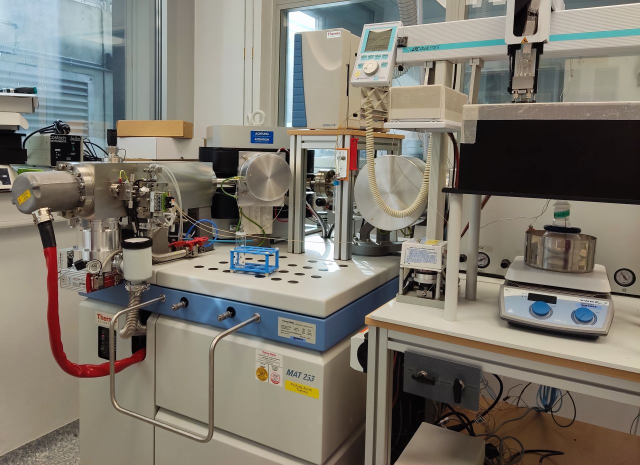
CF-IRMS stands for Continuous Flow Isotope Ratio Mass Spectrometry. It is an analytical method from isotope geochemistry and analytical chemistry that enables the ratio of stable isotopes of an element in a sample to be determined very precisely, for example carbon (13C/12C), nitrogen (15N/14N) or oxygen (18O/16O).
Contact person: Prof. Dr. B. Schöne, Michael Maus; – Research group Paleontology
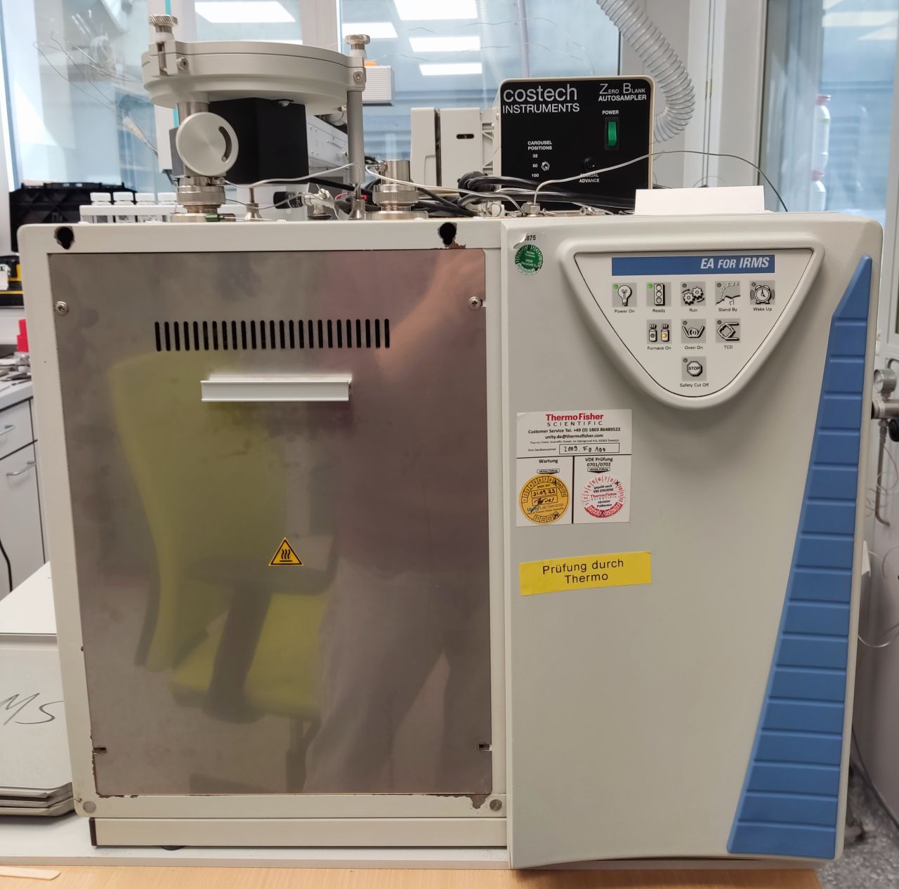
TC-EA stands for Thermal Combustion Elemental Analyzer. It is used to reduce various oxygen-containing substances such as phosphates, waters, or cellulose to carbon monoxide at high temperatures of 1450°C in a glassy carbon reactor, the oxygen isotope composition of which is then measured in a CF-IRMS.
Contact person: Prof. Dr. B. Schöne, Michael Maus – Research group Paleontology
EA stands for Elemental Analyzer. With this device, organic substances such as soil, leaves, hair, collagen, cellulose, or biominerals weighed into tin capsules are oxidized at 1050°C in a cobalt oxide-containing reactor with the addition of oxygen. A downstream reactor with elemental copper eliminates excess oxygen or reduces nitrogen oxides to nitrogen gas. Using a connected CF-IRMS, the isotope ratios of stable isotopes of carbon (13C/12C), nitrogen (15N/14N), or sulfur (34S/32S) in CO2 or N2 can be determined with an accuracy of a few tenths of a per mil.
Contact person: Prof. Dr. B. Schöne, Michael Maus – Research group Paleontology
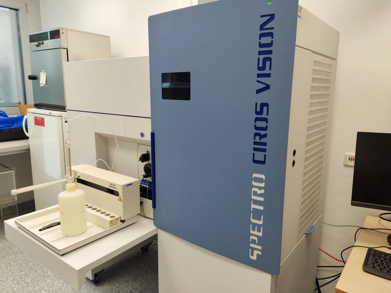
ICP-OES is an analytical method from analytical chemistry for determining the elemental composition of a sample. In an Ar plasma, the atoms of the elements are excited and emit characteristic light radiation. This emitted radiation is separated by wavelength and measured in an optical emission spectrometer. Since each element has a typical emission spectrum, the elements present can be identified and their concentrations determined. ICP-OES is frequently used in environmental analysis, materials science, geochemistry, and food analysis to quickly and simultaneously determine metals and trace elements in a sample.
Contact person: Prof. Dr. B. Schöne, Prof. Dr. Thomas Tütken – Research group Paleontology
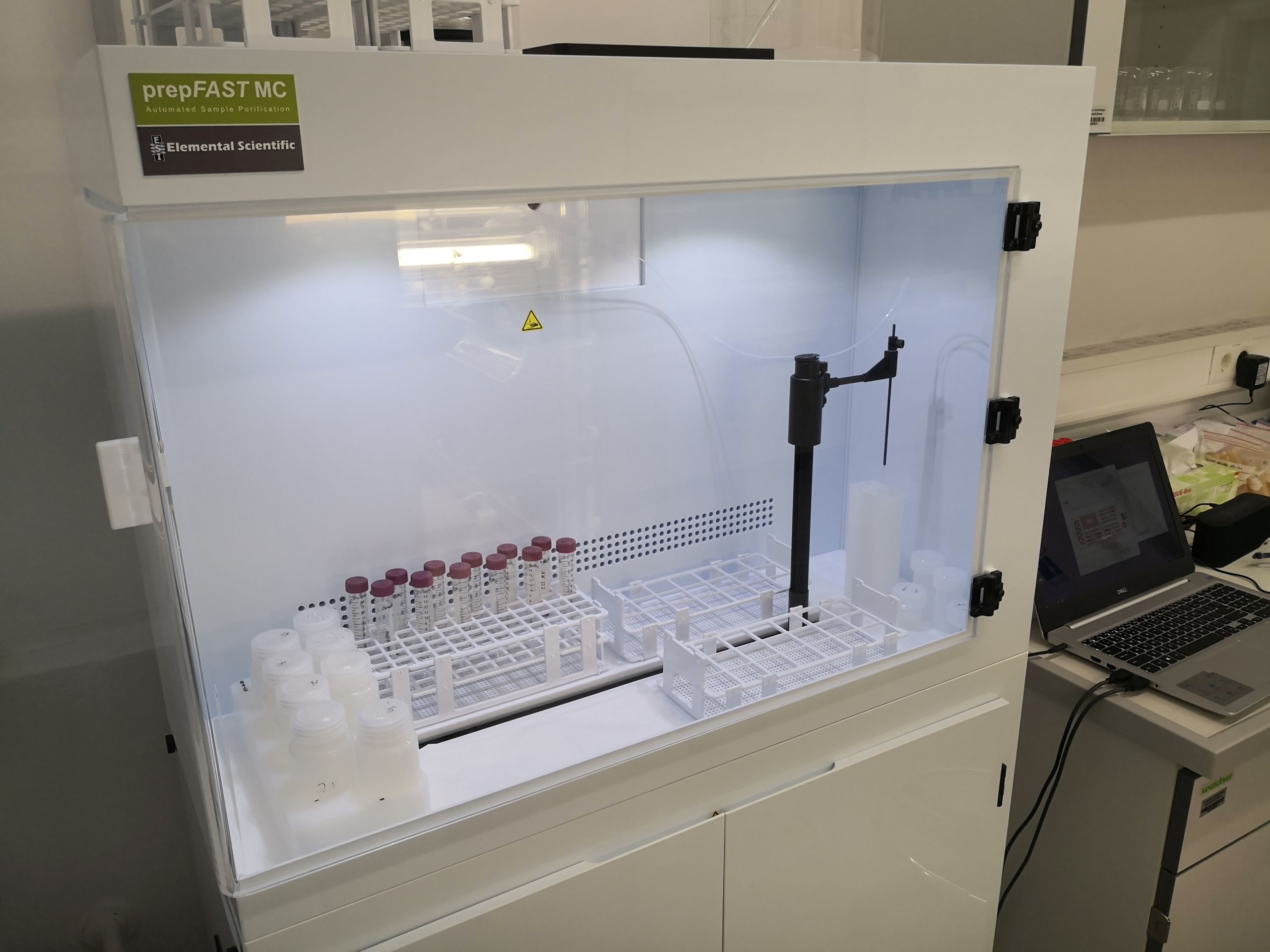
PrepFAST from Elemental Scientific (ESI) is used to automate sample preparation for MC-ICP-MS isotope analysis of heavy isotopes. It enables automated ion-chromatographic separation of the elements Ca and Sr from the matrix for isotope analysis. For this purpose, the sample must first be dissolved in HCl.
Contact person: Prof. Dr. Thomas Tütken – Research group Paleontology
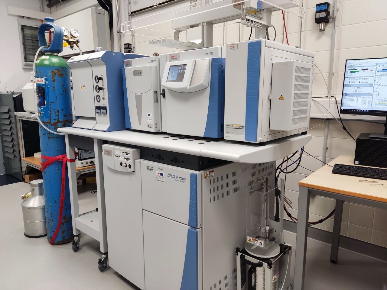
GC-C-IRMS (gas chromatography–combustion–isotope ratio mass spectrometry) is an analytical method for determining stable isotope ratios of individual compounds within a sample. With this technique, the individual components of a sample are first separated from one another using a gas chromatograph (GC). The separated compounds are then converted into simple gases such as CO₂ or N₂ in a combustion furnace (combustion interface). These gases are subsequently analyzed in an isotope ratio mass spectrometer (IRMS), which determines the ratios of stable isotopes (e.g., ¹³C/¹²C or ¹⁵N/¹⁴N) with high precision. The method is used in various scientific fields, including environmental research, food analysis, forensics, and geochemistry. In particular, it is used to investigate sources, origin, and biogeochemical processes of organic compounds.
Contact person: Prof. Dr. B. Schöne – Research group Paleontology
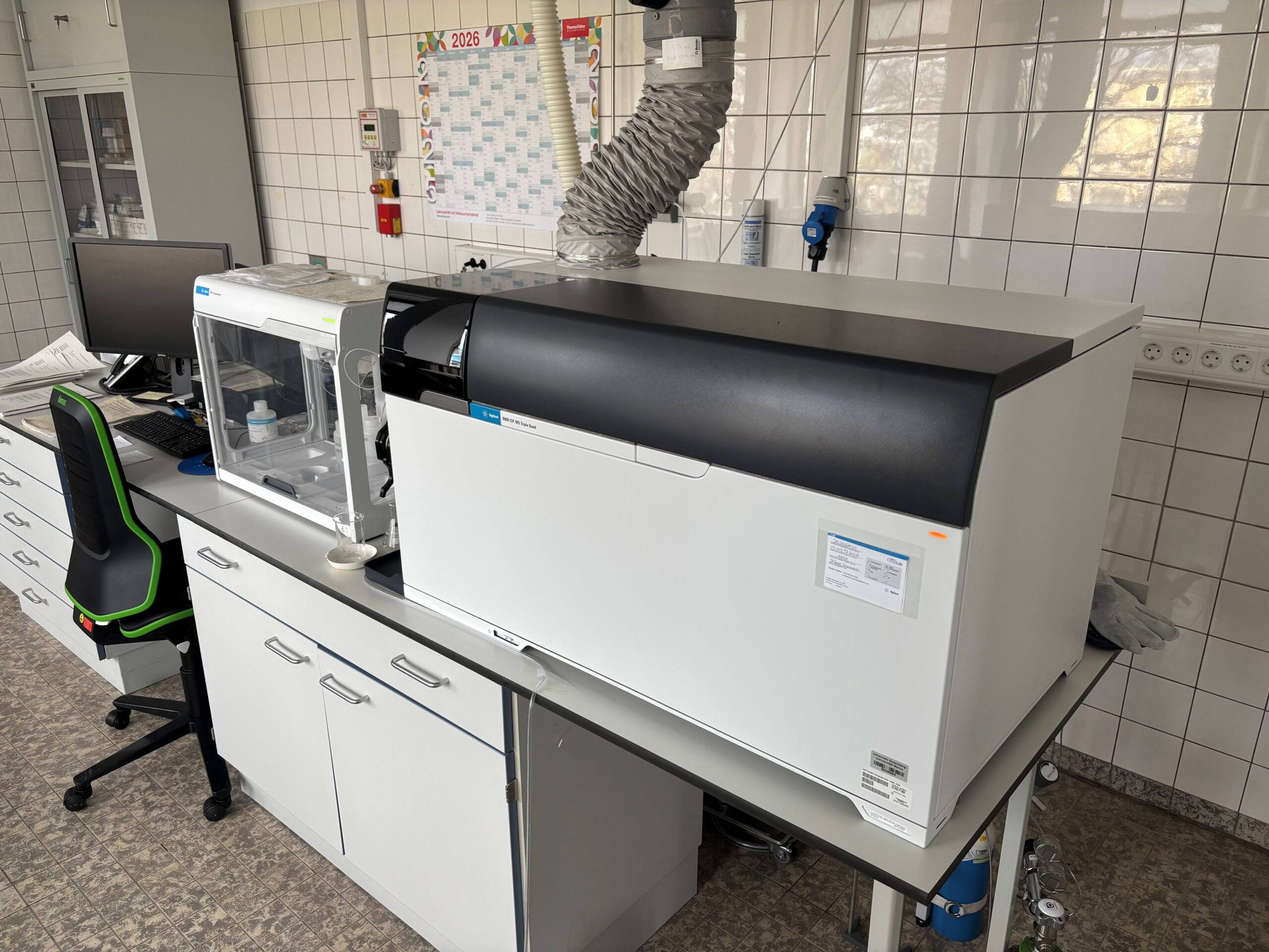
The Triple-Quad ICP-MS (inductively coupled plasma mass spectrometry) is used to determine element concentrations in dissolved and leached rocks and waters down to the ppt (parts per trillion) range.
Contact person: Prof. Dr. Philip Pogge von Strandmann – Research group Sediment Geochemistry
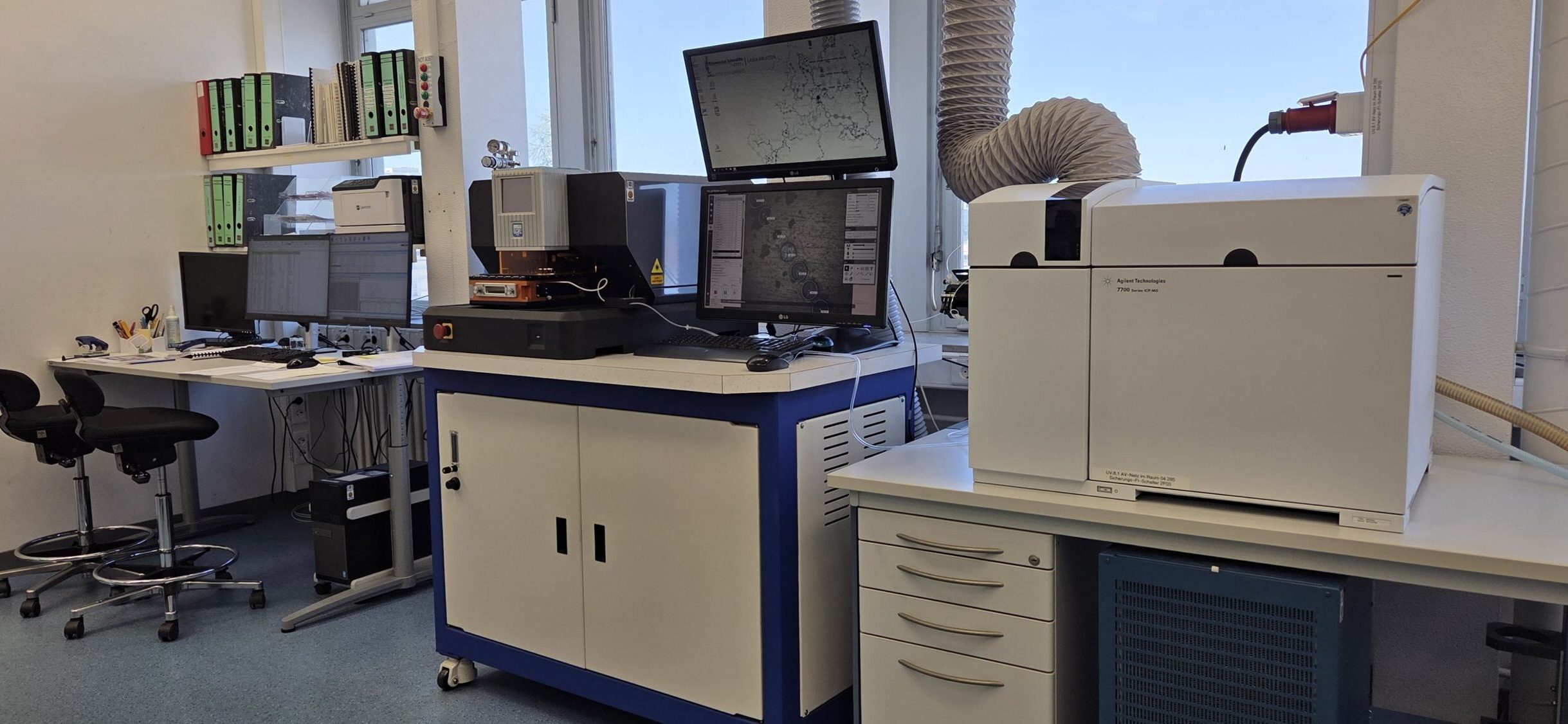
LA-ICP-MS (“Laser Ablation – Inductively Coupled Plasma – Mass Spectrometry”) is a sensitive analytical method for rapid multi-element determination in the trace and ultra-trace range in solid sample materials and technical products. The sample material to be examined is ablated using focused laser radiation and transported with a carrier gas (argon or helium) into the inductively coupled plasma ion source of the ICP-MS. In the plasma at approx. 8000°C, the tiny sample particles are atomized and positively ionized, accelerated, and transported into the high vacuum of a mass spectrometer. There, they are separated according to their mass-to-charge ratio and energy-to-charge ratio and detected with time resolution. The resulting laser craters are only a few µm in size; therefore, this minimally invasive method is also well suited for valuable material (museum objects, gemstones). In the geosciences laboratory at the university of Mainz, LA-ICP-MS has been used since 2004 in trace element analytics to characterize samples from the fields of geology, Mineralogy, climate research, materials science, gemstone research, archaeology, and isotope analytics, e.g., for U-Pb dating of zircons.
Contact person: Dr. Regina Mertz, Prof. Dr. Denis Scholz – Research group Speleothem Research
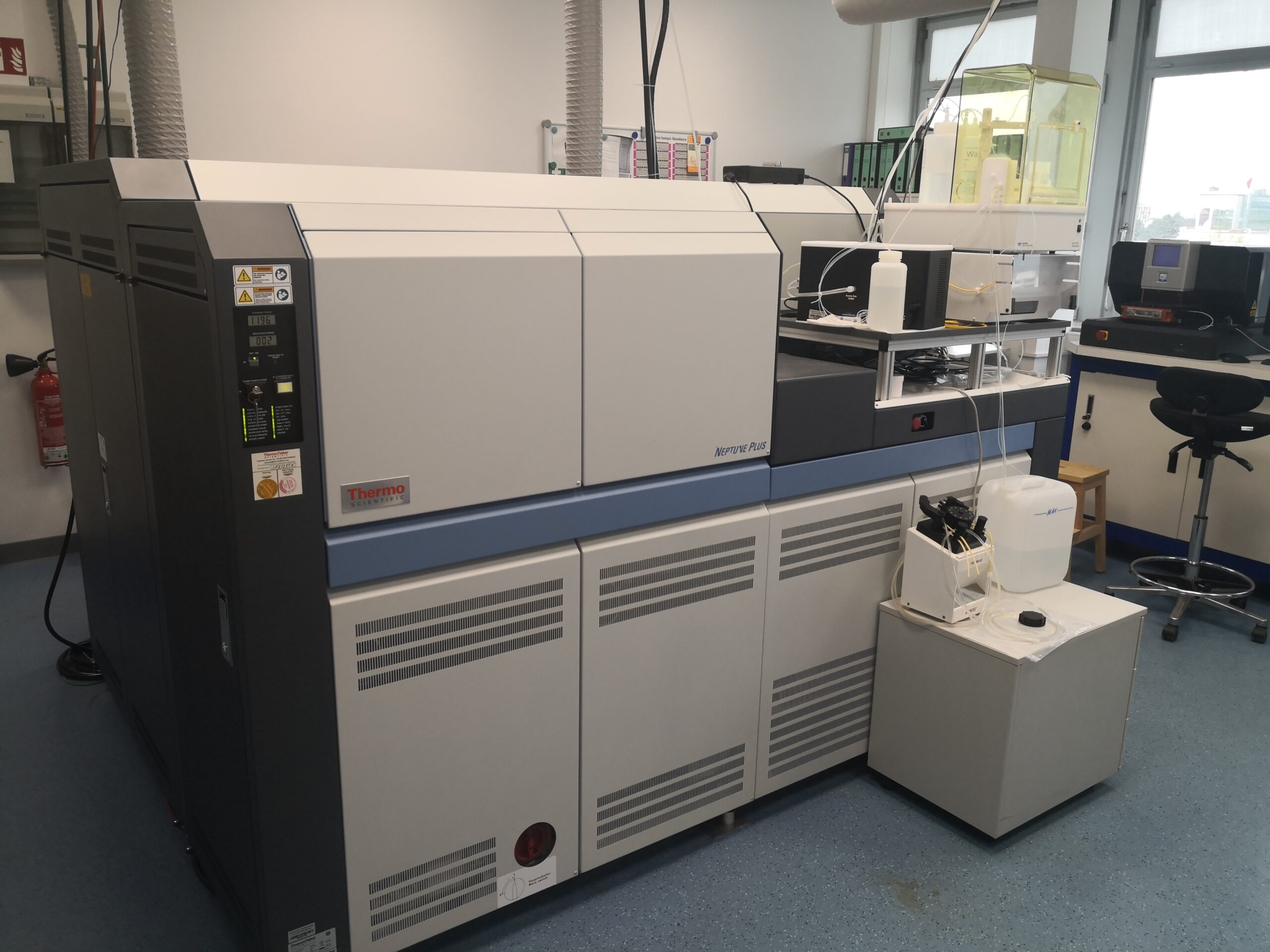
MC-ICP-MS (“Multi Collector – Inductively Coupled Plasma – Mass Spectrometry”) is a highly sensitive analytical method for the precise determination of isotope ratios in the trace and ultra-trace range in solid and liquid sample materials. The sample material to be analysed is transported either directly via laser ablation or, after prior wet-chemical preparation in the cleanroom laboratory, with a carrier gas into the inductively coupled plasma ion source of the ICP-MS. In the plasma, which is approx. 8000°C hot, the tiny sample particles are atomised and positively ionised, accelerated, and transported into the high vacuum of the mass spectrometer. There, they are separated according to their energy-to-charge ratio and mass-to-charge ratio and simultaneously detected with time resolution in several detectors of differing sensitivity.
In our laboratory, we have been measuring various isotope systems on different geoscientific sample materials since 2019. One focus is high-precision dating of carbonates over the last 650,000 years using the 230Th/U method, including in-situ via laser ablation. We also focus on the following isotope systems: strontium, calcium, magnesium, and lithium.
Contact person: Prof. Dr. Denis Scholz, Dr. Regina Mertz, Dr. Michael Weber – Research group Speleothem Research
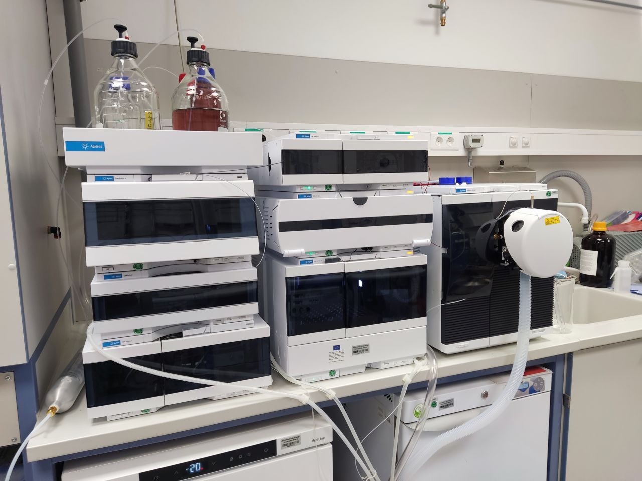
LC-MS with electrospray ionization (ESI) is a powerful analytical method for investigating and determining organic compounds in complex samples. First, the components of the sample are separated from one another by a liquid chromatograph (LC). The eluting compounds then enter an electrospray ionization source, where a fine, electrically charged
Contact person: Prof. Dr. B. Schöne – Research group Paleontology
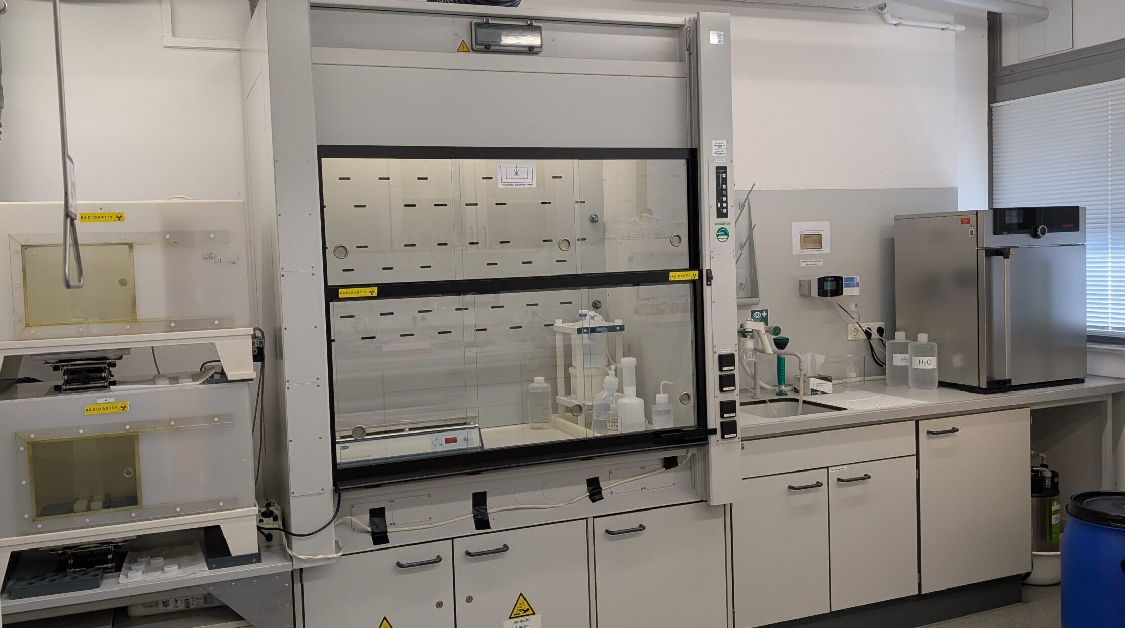
The metal-free cleanroom laboratory is used for the preparation and purification of samples for metal isotope analysis.
Contact person: Prof. Dr. Philip Pogge von Strandmann – Research group Sediment Geochemistry
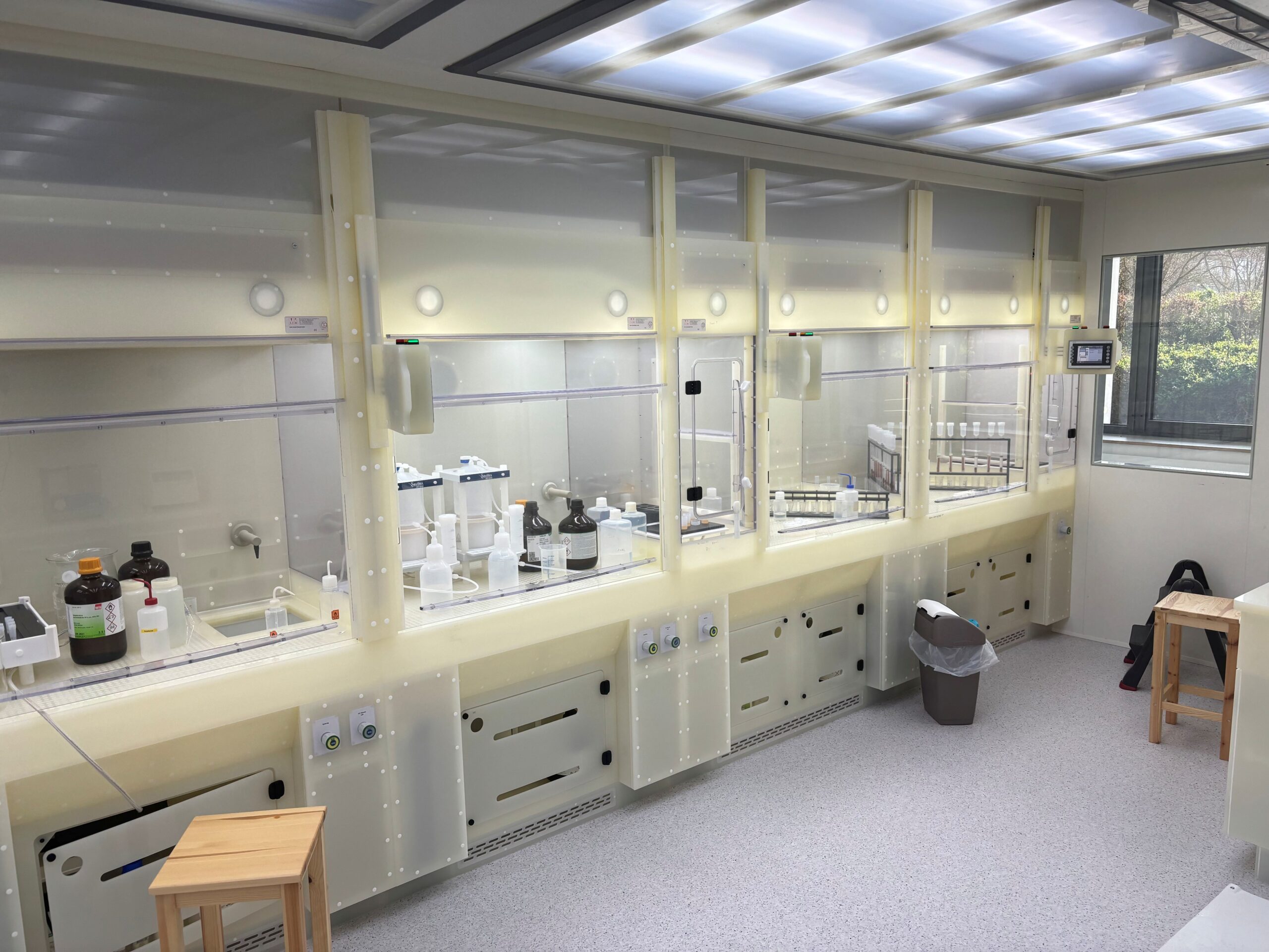
In our cleanroom laboratory, we carry out sample preparation for mass spectrometric analysis. Using ion-exchange column chromatography, the elements to be analysed are separated from the sample matrix (carbonate, gypsum, phosphates, aqueous solutions).
Contact person: Prof. Dr. Denis Scholz, Susann Denndorf – Research group Speleothem Research
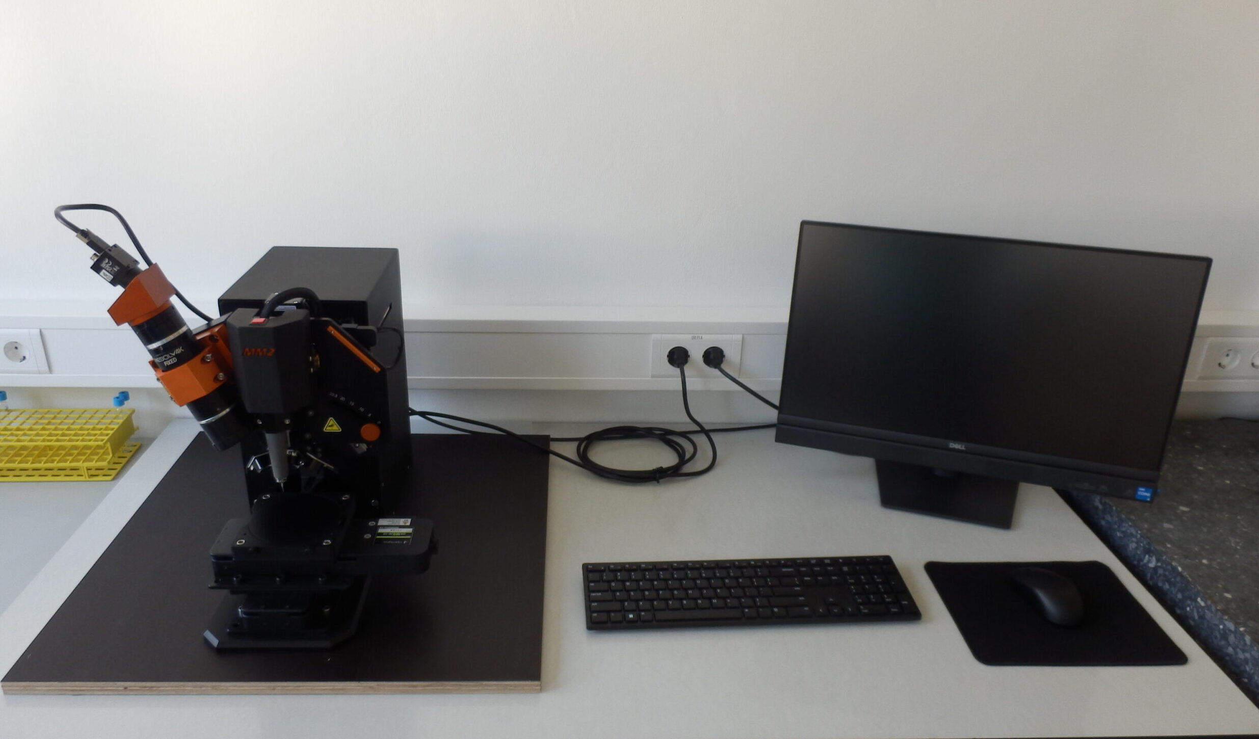
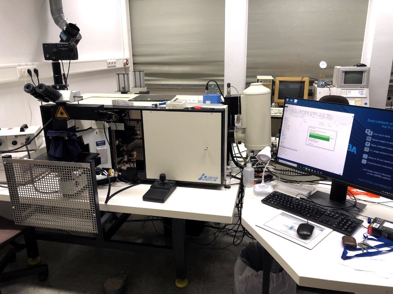
Confocal micro-Raman spectrometer, equipped with four possible excitation lasers (blue: 488 nm, green: 532 nm, red: 633 nm, infrared: 785 nm) and three possible diffraction gratings (300 gr/mm, 900 gr/mm, 1800 gr/mm)
Contact person: Dr. Tobias Häger – Research group Geomaterials and Gemstone Research
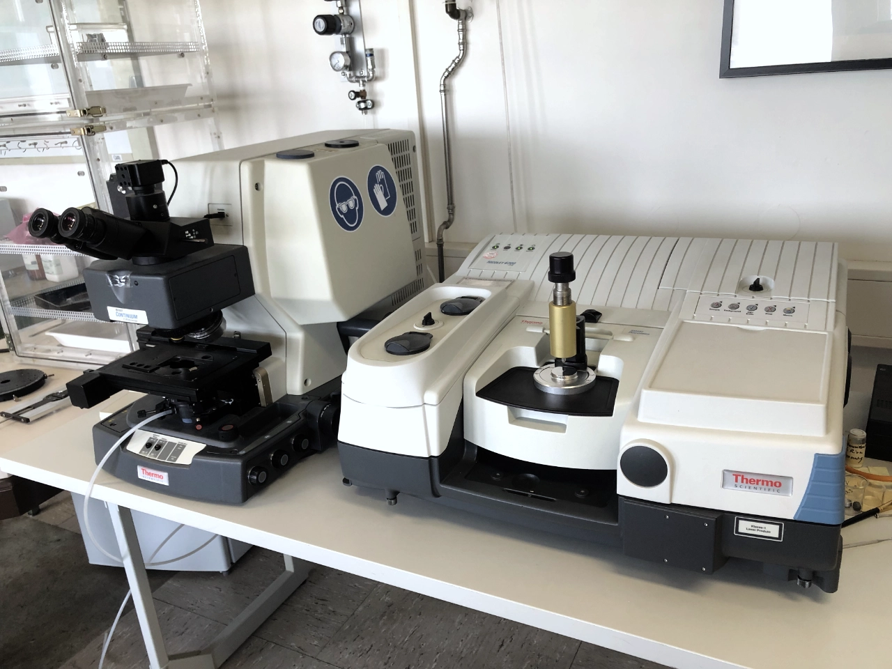
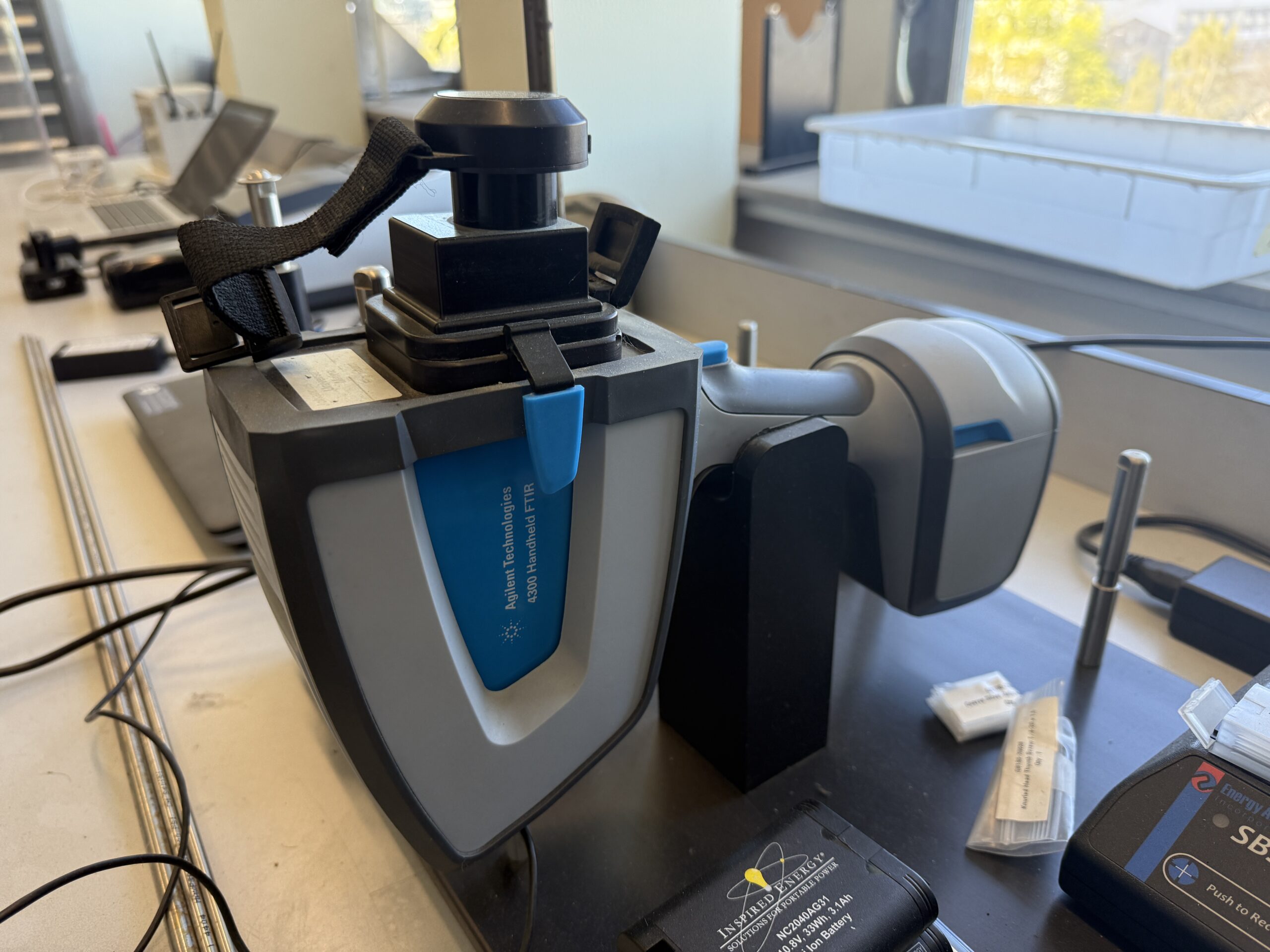
Fourier transform infrared (FT-IR) spectrometer with connected microscope unit for IR absorption analysis of powder samples and mineral thin sections in the near, mid and far infrared range.
Contact person: Dr. Tobias Häger – Research group Geomaterials and Gemstone Research
UV-Vis-NIR Microspectrophotometer. Two spectrometers (TIDAS CCD UV/NIR and TIDAS PSS NIR) are connected to the Zeiss Axio Imager.A2m, covering the complete range from 300nm-1600nm. Transmission measurements in the 0.1mm range are possible.
Contact person: Dr. Tobias Häger – Research group Geomaterials and Gemstone Research
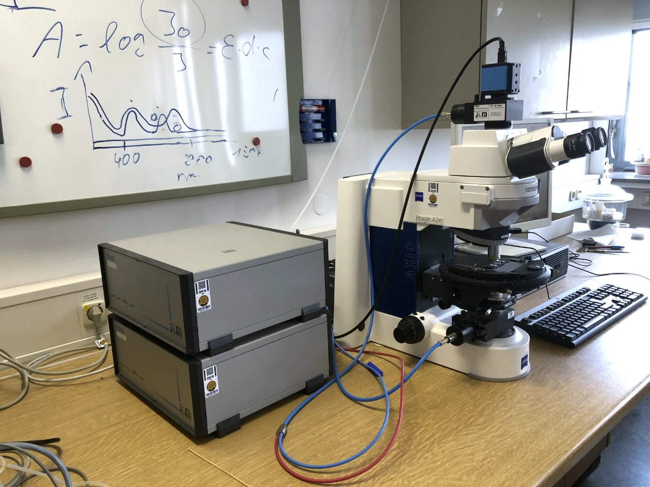
UV/Vis Spectroscopy is used to determine and quantify substances in solution based on their light absorption in the ultraviolet and visible wavelength range. A sample is irradiated with light in the UV and visible range. Certain molecules absorb light at characteristic wavelengths, causing electrons to be excited to higher energy levels. The attenuation of light intensity is measured and can be used to determine the concentration of a substance. Application areas include chemical analysis, environmental analysis, food analysis, and biochemical research, particularly for the rapid and simple determination of dissolved substances such as proteins, nucleic acids, dyes, or metal ions.
Contact person: Prof. Dr. B. Schöne, Michael Maus – Research group Paleontology

Contact person: Prof. Dr. J. Castro – Research group Volcanology
The Specim Hyperspectral Imaging systems provide high-resolution, non-destructive analysis of sediments, rocks and other materials across a broad spectrum. With VNIR (Visible to Near-Infrared, 400–1000 nm) and SWIR (Short-Wave Infrared, 1000–2500 nm) cameras, both visible and infrared properties of the samples can be captured.
This technology enables rapid acquisition of 2D spectral data, allowing conclusions to be drawn about minerals, organic matter, pigments, particle size and sedimentary structures. By combining VNIR and SWIR data, researchers can precisely identify and spatially visualise different sediment components, making it an important tool for high-resolution geoscientific analyses.
Contact person: Jun.-Prof. Dr. I. Obreht – Research group High-Resolution Sedimentology
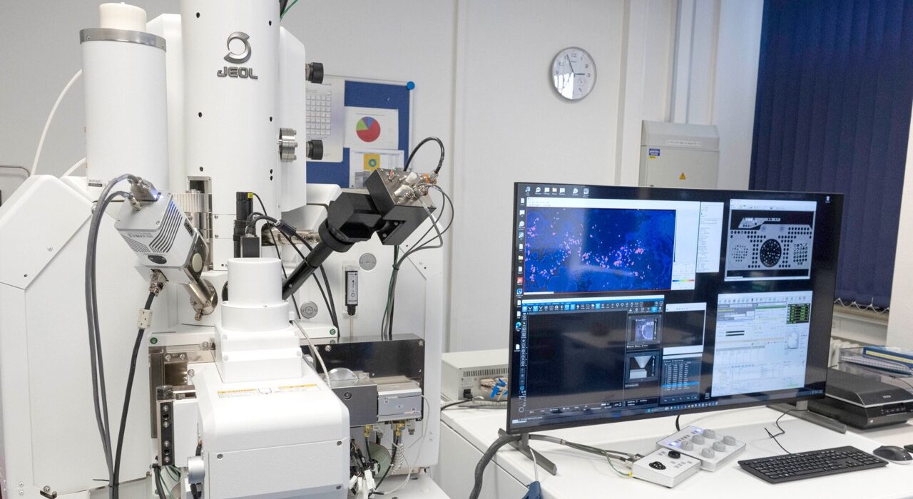
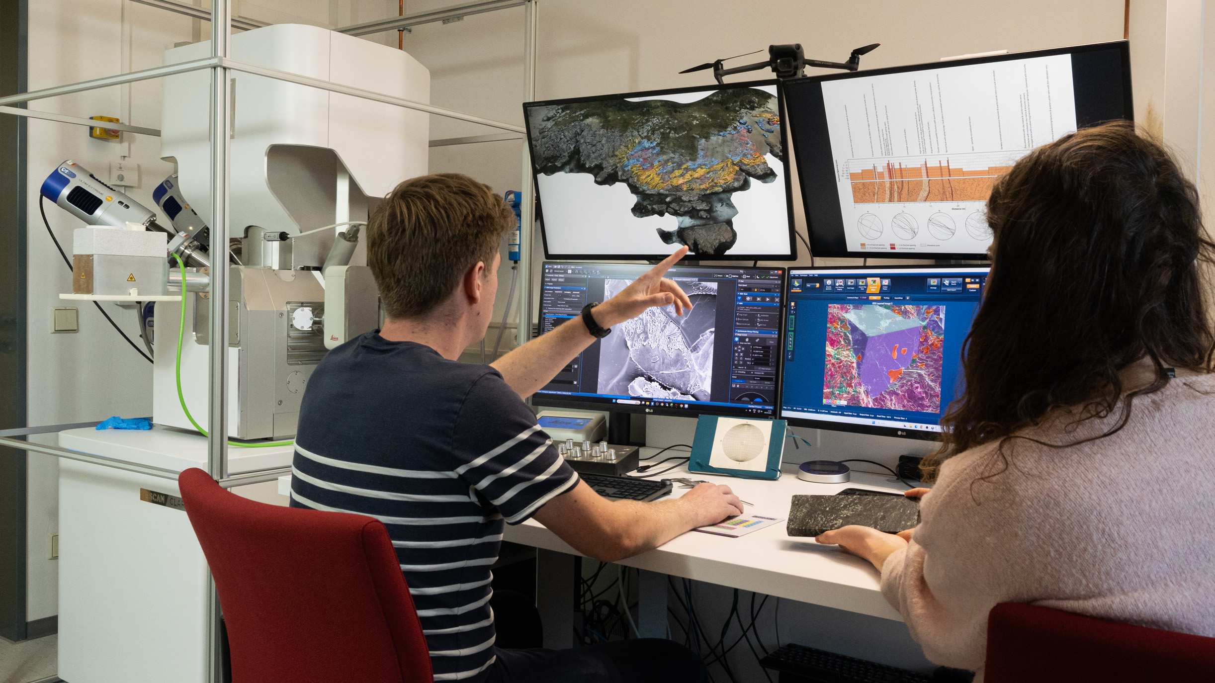
Our scanning electron microscope is equipped with:
- LE-BSE high-contrast backscatter detector, specifically for low acceleration voltage and thus higher resolution
- ET secondary electron detector
- In-lens SE and BSE for high resolution
- EDS “Oxford Ultim Max”
- EDS “Oxford Ultim Extreme”, windowless EDS detector for light elements and low accelerating voltage
- EBSD “Oxford Symmetry 3”
- Cathodoluminescence detector
Contact person: Friedrich Hawemann – Electron Microscopy Centre Mainz
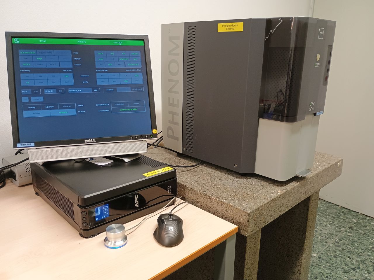
The 3rd-generation Phenom PRO offers powerful imaging capabilities in a compact desktop format. It is optimized for users who require precise results with maximum throughput. The ProSuite platform extends the Phenom PRO with specialized analysis tools to extract quantitative data directly from SEM images. In addition, the Leica EM ACE200 sample coater enables fully automated sputter coating with platinum (Pt) and gold/palladium (Au/Pd) for optimal conductivity.
- Analysis: Voltage levels of 5 kV and 10 kV to optimize material contrast and surface details.
- Imaging: Resolution up to 17 nm at magnifications from 20x to 100,000x.
- Electron source: High-intensity CeB6 source for increased brightness and detail resolution.
- Workflow: Sample exchange and image display in 30 seconds.
- Navigation: Combined optical color camera and electron optics for intuitive sample positioning.
Contact person: Prof. Dr. B. Schöne – Research group Paleontology
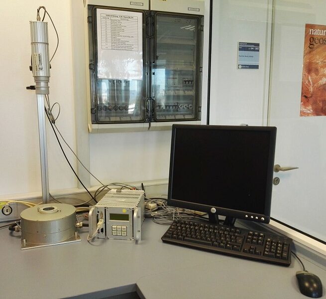
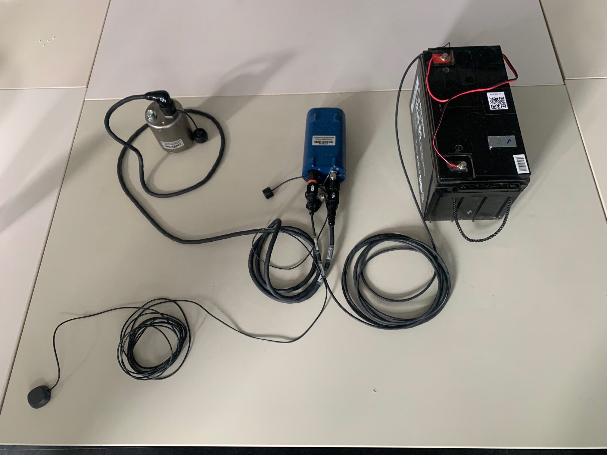
Compact broadband stations are used for many applications in modern seismology. The Volcanic Seismology work group has acquired 16 seismic stations consisting of a Nanometrics Trillium Compact seismometer combined with data loggers such as Pegasus andCentaur. The Trillium Compact is a high-resolution broadband seismometer that enables precise recording of ground motion thanks to its wide frequency range (typically 120 s–100 Hz), low self-noise, and high sensitivity.
Thanks to its compact design, low power consumption, and easy installation, the sensor is suitable both for temporary field experiments, including direct burial or operation in humid environments, and for permanent measurement stations.
Data acquisition is carried out using modern, energy-efficient data loggers. The Pegasus logger is designed in particular for portable, quickly deployable stations and enables autonomous operation with very low power consumption, while the Centaur logger is used as a more powerful alternative with an extended range of functions, a higher channel count, and integrated networking and real-time data transmission.
Together, these components form a robust, flexible, high-resolution measurement station that is suitable both for temporary experiments with high station density and for long-term monitoring networks.
Contact person: Jun.-Prof. Dr. M. Reiss – Research group Volcano Seismology
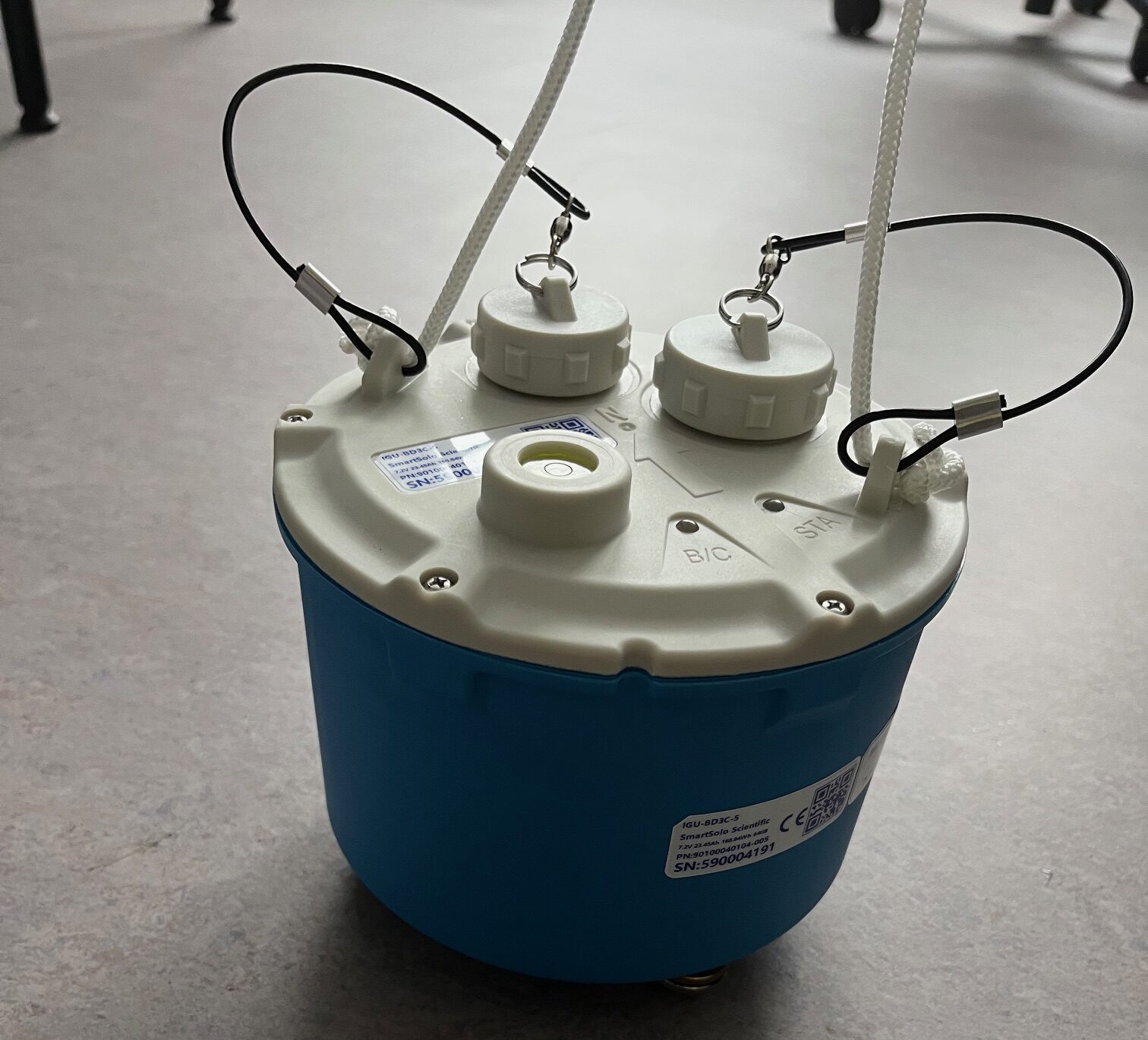
Seismic nodes are compact, self-contained instruments used for temporary recording of ground motion. Unlike traditional seismic stations, they combine the sensor, data storage, power supply, and time reference in a single, usually rugged housing. Due to their small size and easy installation, they can be deployed in large numbers and at high spatial density.
They are typically used for time-limited experiments, for example for high-resolution imaging of subsurface structure or for detailed investigation of seismic activity. After the measurement campaign is completed, the devices are collected again and the recorded data are read out and analysed.
Contact person: Jun.-Prof. Dr. M. Reiss – Research group Volcano Seismology
Simultaneous thermogravimetry and dynamic differential calorimetry
Contact person: Dr. Tobias Häger – Research group Geomaterials and Gemstone Research
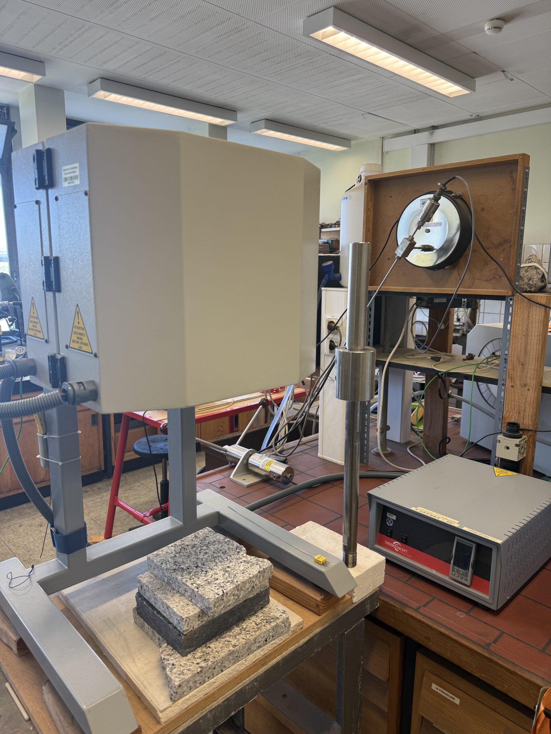
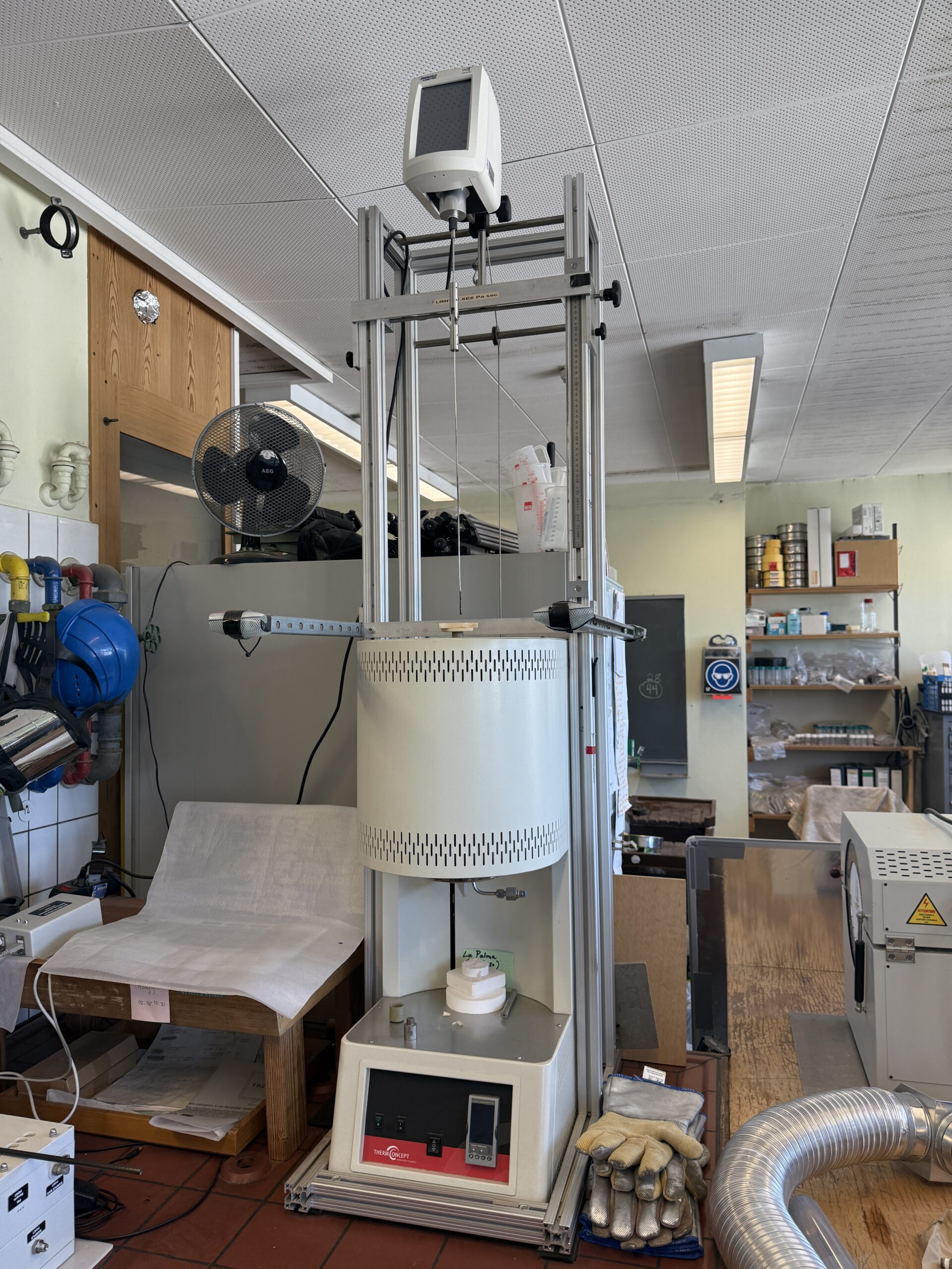
Various high-temperature furnaces, in some cases up to 1800°C
Contact persons: Prof. Dr. R. Botcharnikov, Dr. S. Buhre, Prof. Dr. J. Castro, Dr. T. Häger
In the Biomineralisation research group, photosynthetic microalgae are cultivated in both marine and freshwater under controlled conditions, with a particular focus on dinoflagellates. The current collection includes 33 species cultivated in 10 media.
Taxonomically, dinoflagellates (13 strains) dominate, supplemented by diatoms (7), green algae (5), cryptophyceae (3), cyanobacteria (2), synurales (2), and haptophytes.
Contact person: Jun.-Prof. Dr. A. Jantschke – Research group biomineralization
The PLG 400 is a climate chamber for the controlled cultivation of plants and microorganisms under defined environmental conditions. Temperature, light intensity, and day/night cycles can be precisely adjusted to ensure reproducible growth conditions.
Contact person: Jun.-Prof. Dr. A. Jantschke – Research group biomineralization
For sterile work, an autoclave (Systec V-95) and a laminar flow workbench (SafeFAST Classic) are used. The autoclave enables the sterilization of media, solutions, and equipment using saturated steam under pressure. The sterile bench ensures a particle-free working environment through HEPA-filtered laminar flow, allowing cultures and samples to be processed under aseptic conditions.
Contact person: Jun.-Prof. Dr. A. Jantschke – Research group biomineralization
The Olympus IX70 is an inverted fluorescence microscope for examining samples in transmitted light and epifluorescence modes. Due to its inverted design, it is particularly suitable for the analysis of samples in liquids or cell cultures. The system features modular filter cubes for epifluorescence and the possibility to integrate polarization filters, allowing for the investigation of anisotropic material properties in addition to fluorescently labeled structures.
Contact person: Jun.-Prof. Dr. A. Jantschke – Research group biomineralization
The LUNA-FL is an automated fluorescence cell counter from Logos Biosystems for rapid and reproducible determination of cell count and cell viability. The system combines microscopic image acquisition with integrated software analysis and enables both brightfield and fluorescence measurements.
Contact person: Jun.-Prof. Dr. A. Jantschke – Research group biomineralization
The automatic titrator Mettler Toledo DL58 is a microprocessor-controlled analysis system for precise and reproducible execution of various titrations. It enables, among other things, potentiometric, photometric, amperometric, and pH-stat titrations and offers flexible method creation through freely programmable procedures.
In the Biomin research group, the system is used for the controlled synthesis of amorphous calcium carbonate (ACC) to precisely control reaction parameters such as pH value and titration rate. Through automated, incremental addition of reagents and continuous monitoring of the solution (e.g., pH or potential), supersaturation and nucleation can be specifically regulated, enabling reproducible and controlled ACC formation.
Contact person: Jun.-Prof. Dr. A. Jantschke, C. Liedgens – Research group biomineralization